We are dedicated to pushing the edge of computation and efficiency in healthcare AI.
I work on statistical machine learning (ML), healthcare big data system and applications in medical image analysis and biomedicine. I'm currently working as a research graduate student and pursuing a Ph.D. My supervisor is Dr. Bennett A. Landman, who perform exterme enthusiuasm in translational research to explore innovative, clinical useful techniques. Our group develops new science and technologies that make machine learning and deep learning (DL) more robust/reliable and reveals clinical insights.
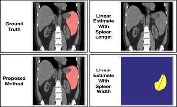
Our team has close collaboration and advise with Vanderbilt University Medical Center (VUMC) and industry groups. We investigate new, interesting hypotheses ranging from brain connectomics, optic nerve disease, lung cancer to abdominal metabolic syndrome and renal drainage. My methodological research mainly focuses on abdominal imaging, metabolic syndrome, renal structures and lung cancer prediction. Interests but not main works for exploring brain connectomics and infrastructures in medical imaging big-data system.
(Below are some of my active research interests areas, please refer to my Google Scholar for full research scopes in the institutional collaborators)
MONAI: Open Healthcare AI Infrastructure
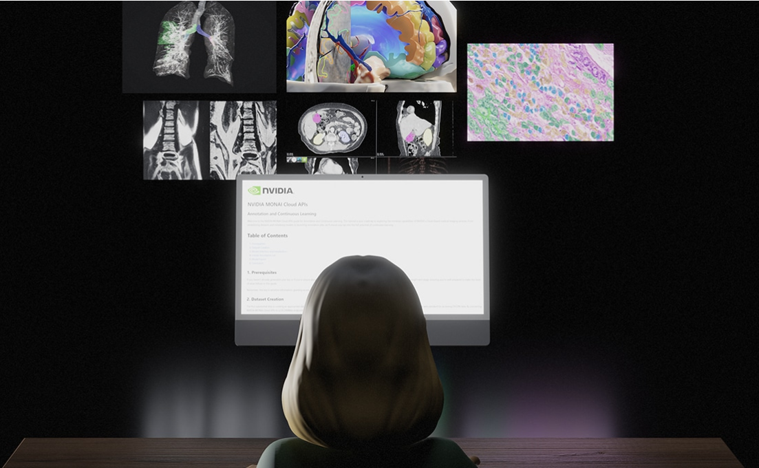
With NVIDIA MONAI cloud APIs, solution providers can more easily integrate AI into their medical imaging platforms, enabling them to provide supercharged tools for radiologists, researchers and clinical trial teams to build domain-specialized AI factories. The APIs are available in Nvidia Cloud Foundation ecosystem through the NVIDIA DGX Cloud AI supercomputing service.
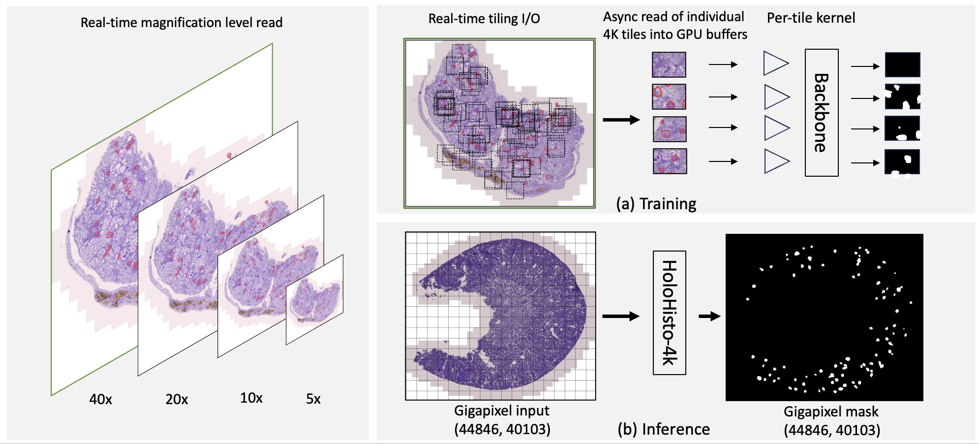
HoloHisto: Holistic WSI computing Framework
We developed an end-to-end workflow for training and inferencing gigascale WSI, introducing a novel learning paradigm to the field of WSI analysis.. HoloHisto is designed to handle inputs and outputs of any size, regardless of whether they are (WSIs) or smaller patches. To assess the performance, our team curated a new kidney pathology image segmen- tation (KPIs) dataset with WSI-level glomeruli segmentation from whole mouse kidneys. From the results, HoloHisto-4K delivers remarkable performance gains over previous state-of-the-art models. The source code HoloHisto platform and the KPIs dataset is publicly available as open-source to advocate re- producible research for the community.
Robust/reliable ML/DL and Statistics
We have led research in robust deep learning algorithms (e.g. outlier-robust learning via meta parameterization, active learning). We also developed methods on reducing human efforts in automatic labeling (e.g. semi-supervised learning via quality assesments, partially-supervised segmentation and weakly-supervised labeling via self-supervision). Our methods are used in daily basis.
Renal & Pancreatic Imaging
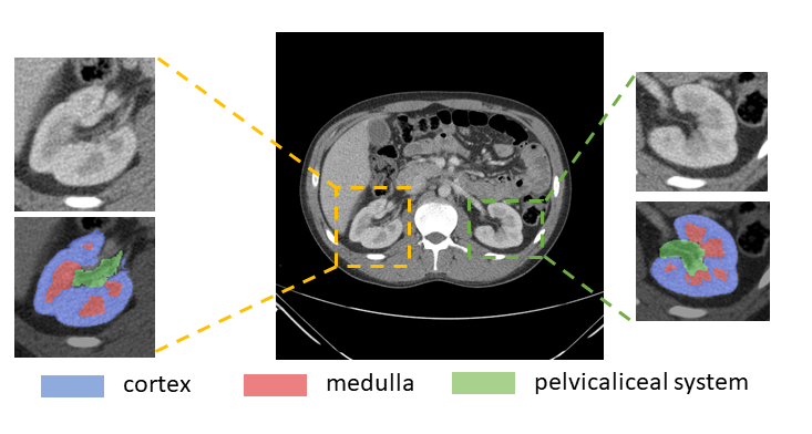
We created one of the world-first renal segmentation dataset with renal cortex, medulla and pelvicalyceal system. We have radiologists labeled 102 subjects on contrast enhanced CT scans. We developed coarse to fine labeling paradigms so that automated methods can be involved to enable efficient labeling (Radiology: Artificial Intelligence). We perform to identify volumetric biomarkers on renal structures to assist of prognosis, guide treatment selection, treatment deliveries. We also created paried CT-MRI pancreatic studies for evaluting human pancreas and diabetes, pancreatitis context, including but not limit to contruction of atlas, segmentation of pancreas under context, pancreatic ducts in different modalities.
Abdominal Imaging
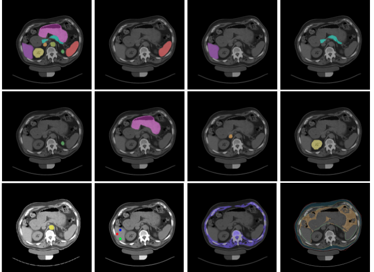
We have developed and evaluated algorithms for the automated labeling of abdominal structures (segmentation) in patient populations using current generation clinically acquired CT data. We released our challenge dataset in MICCAI2015 Multi-Atlas Labeling Beyond the Cranial Vault. We have created new labeling paradigms so that automated methods can be efficiently learned from expertly labeled training data (MICCAI20, MedIA). We perform to identify biomarkers based on structural imaging to improve accuracy of prognosis, guide treatment selection, and improve patient outcomes.
Metabolic Syndrome
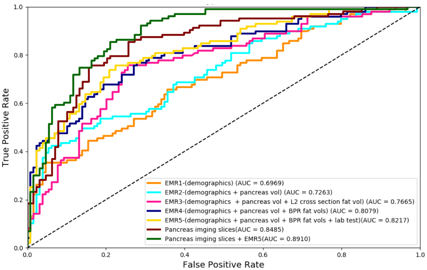
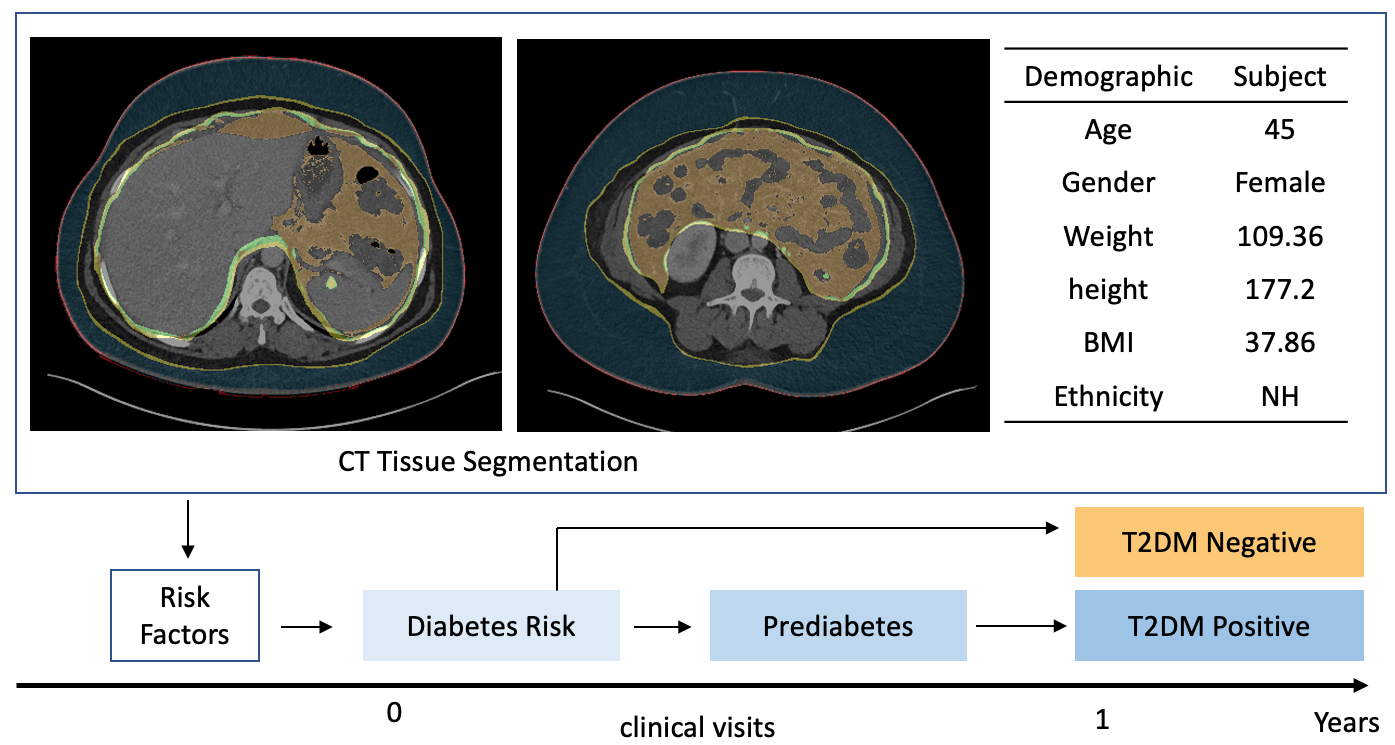
Type II diabetes mellitus (T2DM) is a significant public health concern with multiple known risk factors (e.g., body mass index (BMI), pancreas volume, body fat distribution, glucose levels). Improved prediction or prognosis would enable earlier intervention before possibly irreversible damage has occurred. We explored secondary use of the CT imaging data and the role of different types of electronic medical records (EMR) to refine the risk profile of future diagnosis of type II diabetes. Our metabolic related studies has promising potential for heterogeneous and multi-modal medical applications.
Clinical Biomarkers
Investigations in the imaging structures provide has been used in clinical biomarker associated with disease, infection and cander. We have explored quantitative estimations of biomarkers of clinical interest in molecular, histologic, radiographic characterization of abdominal anatomies (Academic Radiology).Unique challenges emerge when validating machone learning generated imaging biomarkers. We provided changes in imaging hardware, acquisition parameters, and patient positioning during imaging cause significant variations among images.
Lung Cancer Prediction
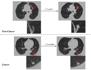
We have studied low dose CTs which have been successful in prediction of future cancer onset. Base on time-to-event analysis, we investigateed cancer accumulating model (MedIA, Riqiang et al.) with clinical risk factors and free-time-to-early-stage-cancer (FTESC) for cancer, non-cancer patients. We also led research in atlas space for lung imaging data, component analysis, latent feature space editing. As the theory-guided data-driven research, our algorithms are used and extended to longitudinal medical applications in Vanderbilt hospital.
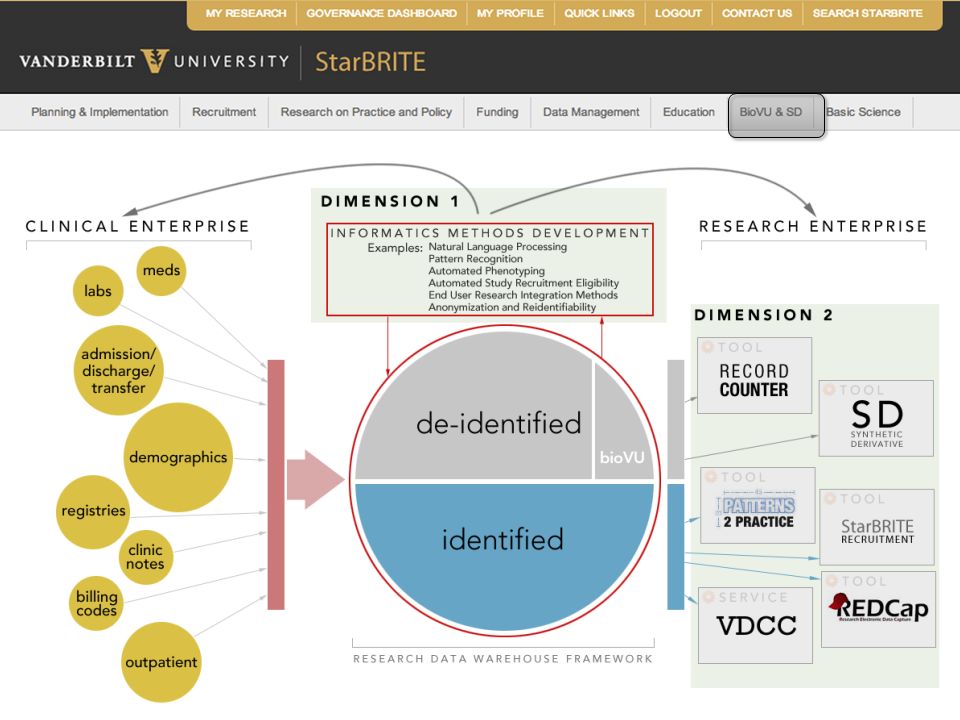
Infrastructures in Medical Big Data
We have deep collaboration research with big data and informatics, we helped and built medical image processing at Vanderbilt to handle data archival. Together with Vanderbilt University Institute of Imaging Science (VUIIS), the computational facilities are created. In additional, ImageVU, record counter, XNAT and RedCAP led the integration of imaging resources with the established clinical data reuse efforts. We have research projects for informatics system, distributed computing and high performance computer.